abstract - in research (preclinical)
BACKGROUND:
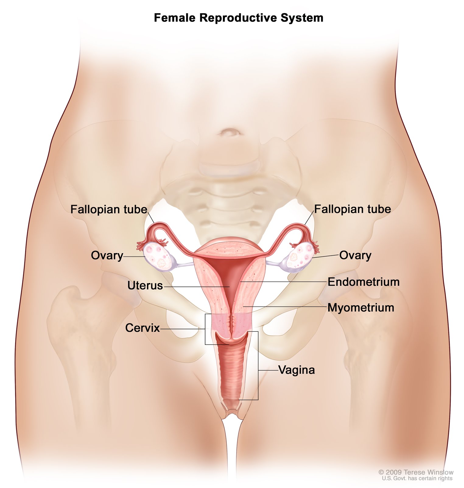
Epithelial
ovarian cancer (EOC) is characterized by the overexpression of cancer
antigen 125 (
CA125), a mucinous glycoprotein that serves as a tumor
biomarker.
Early diagnosis of EOC is plagued by its asymptomatic nature
of progression and the limitations of currently used immunoassay
techniques that detect CA125 as a shed antigen in serum samples.
Presently, there is no technique available for the in vivo evaluation of
CA125 expression in malignant tissues. Moreover, there could be an
unexplored pathophysiological time window for the detection of CA125 in
EOC, during which it is expressed on tumor cells prior to being shed
into the bloodstream. A method for the in vivo evaluation of CA125
expression on ovarian neoplasms earlier along disease progression and/or
recurrence can potentially contribute to better disease management. To
this end, the present work utilizes an anti-CA125 monoclonal antibody
(MAb) and a single-chain variable fragment (scFv) labeled with the
positron-emitting radionuclide (64)Cu for preclinical molecular imaging
of CA125 expression in vivo.
METHODS:
Anti-CA125
MAb and scFv were prepared and functionally characterized for target
binding prior to being tested as radiotracers in a preclinical setting.
RESULTS:
Immunoblotting,
immunofluorescence, and flow cytometry revealed specific binding of
CA125-targeting vectors to NIH:OVCAR-3 cells and no binding to
antigen-negative SKOV3 cells. (64)Cu-labeled anti-CA125 MAb and scFv
were obtained in specific activities of 296 and 122 MBq/mg,
respectively. Both radioimmunoconjugate vectors demonstrated highly
selective binding to NIH:OVCAR-3 cells and virtually no binding to SKOV3
cells. In vivo radiopharmacological evaluation using xenograft mouse
models injected with (64)Cu-labeled anti-CA125 MAb provided a
standardized uptake value (SUV) of 5.76 (29.70 %ID/g) in OVCAR3 tumors
24 h post-injection (p.i.) versus 1.80 (5.91 %ID/g) in SKOV3 tumors.
(64)Cu-labeled anti-CA125 scFv provided an SUV of 0.64 (3.21 %ID/g) in
OVCAR3 tumors 24 h p.i. versus 0.25 (1.49 %ID/g) in SKOV3 tumors.
Results from
small-animal PET imaging were confirmed by ex vivo
autoradiography and immunohistochemistry.
CONCLUSIONS:
Radiolabeling
of anti-CA125 MAb and scFv with (64)Cu did not compromise their
immunoreactivity. Both radioimmunoconjugates presented specific tumor
uptake and expected biological clearance profiles.
This renders them as
potential immuno-PET probes for targeted in vivo molecular imaging of
CA125 in EOC.


0 comments :
Post a Comment
Your comments?
Note: Only a member of this blog may post a comment.