

|
|
|
|
|
|
|
|
|
|
open access
Objective
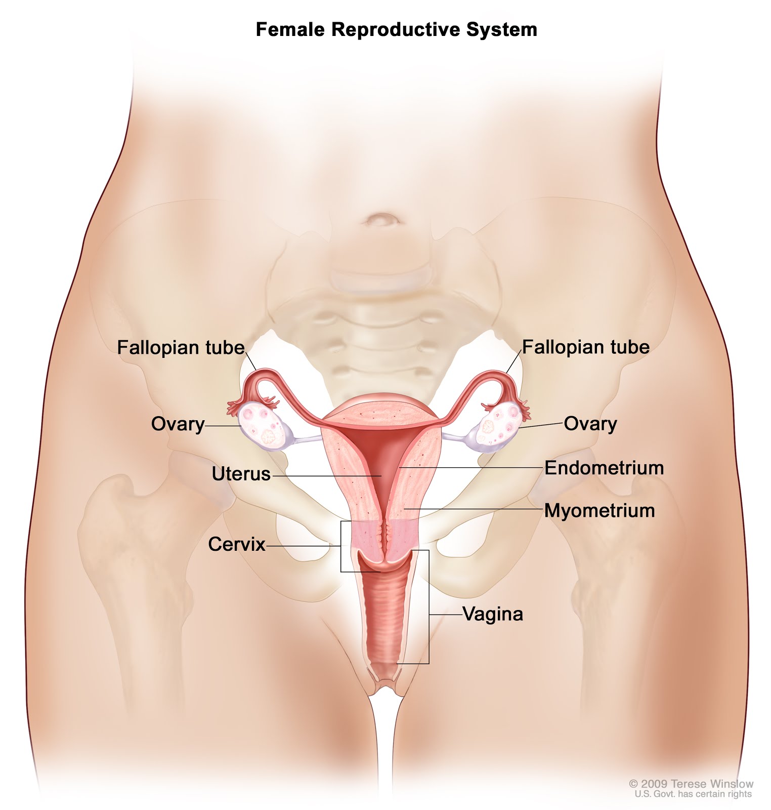
In ovarian cancer, two of the most important prognostic factors for survival are completeness of staging and completeness of cytoreductive surgery. Therefore, intra-operative visualization of tumor lesions is of great importance. Preclinical data already demonstrated tumor visualization in a mouse-model using near-infrared (NIR) fluorescence imaging and indocyanine green (ICG) as a result of enhanced permeability and retention (EPR). The aim of this study was to determine feasibility of intraoperative ovarian cancer metastases imaging using NIR fluorescence imaging and ICG in a clinical setting.
Methods
Ten patients suspected of ovarian cancer scheduled for staging or cytoreductive surgery were included. Patients received 20 mg ICG intravenously after opening the abdominal cavity. The mini-FLARE NIR fluorescence imaging system was used to detect NIR fluorescent lesions.Results
6 out of 10 patients had malignant disease of the ovary or fallopian tube, of which 2 had metastatic disease outside the pelvis. Eight metastatic lesions were detected in these 2 patients, which were all NIR fluorescent. However, 13 non-malignant lesions were also NIR fluorescent, resulting in a false-positive rate of 62%. There was no significant difference in tumor-to-background ratio between malignant and benign lesions (2.0 vs 2.0; P=0.99).Conclusions
This is the first clinical trial demonstrating intraoperative detection of ovarian cancer metastases using NIR fluorescence imaging and ICG. Despite detection of all malignant lesions, a high false-positive rate was observed. Therefore, NIR fluorescence imaging using ICG based on the EPR effect is not satisfactory for the detection of ovarian cancer metastases. The need for tumor-specific intraoperative agents remains.Trial Registration
ISRCTN Registry ISRCTN16945066.....Clinical feasibility trials using this effect with ICG in breast cancer patients in a pre-operative diagnostic setting and in gastric cancer patients during endoscopic surgery showed that it was possible to distinguish tumor from surrounding tissue [18–23]. In addition, Kosaka et al.[24] detected small ovarian (1–2 mm in size) cancer implants using NIR fluorescent imaging after intravenous (IV) administration of ICG in a mouse model. Pathophysiological heterogeneity of solid tumors, for examples in size, presence of necrosis, or presence of vascular mediators may influence accumulation of macromolecules in tumor tissue [25,26]. It is therefore not clear if all preclinical results can be translated to the clinic.


0 comments :
Post a Comment
Your comments?
Note: Only a member of this blog may post a comment.